FFPE Tissue Protein Extraction: A Complete How-To Guide
Formalin-fixed, paraffin-embedded (FFPE) tissue samples are widely used in research and diagnostics due to their preservation of cellular structures, but extracting protein from FFPE tissue can be challenging. This guide covers everything you need to know about FFPE tissue protein extraction, from understanding the importance of various protocols to executing effective methods for extracting high-quality proteins. If you’re navigating the complexities of FFPE protein extraction, this comprehensive guide is here to help.
Understanding FFPE Protein Extraction: Why It’s Essential
FFPE Protein Extraction is a crucial step in many research applications. FFPE samples are preserved by formalin fixation and embedding in paraffin, which stabilizes the protein and maintains tissue morphology over long periods. However, this preservation process cross-links proteins, nucleic acids, and other biomolecules, making them challenging to extract and analyze. Effective protein extraction from FFPE tissue is essential for applications like biomarker discovery, proteomics, and pathology.
FFPE Protein Extraction 3-Step Protocol
Selecting a suitable method is crucial for recovering high-quality proteins when planning your protein extraction protocol. Here, we break down a common protocol for FFPE samples that can be adapted to your research needs.
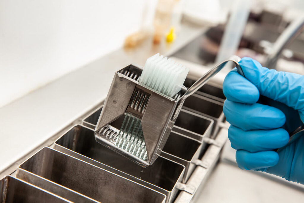
Step 1: Deparaffinization
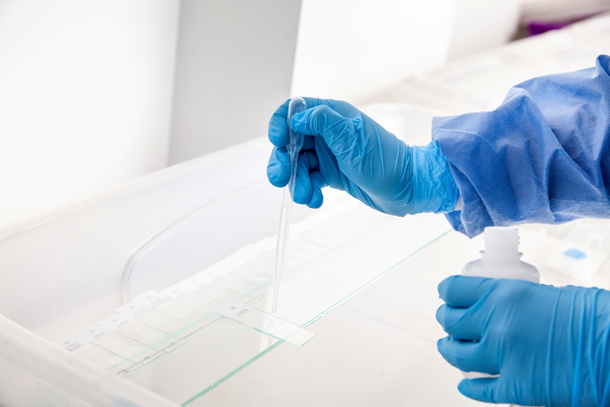
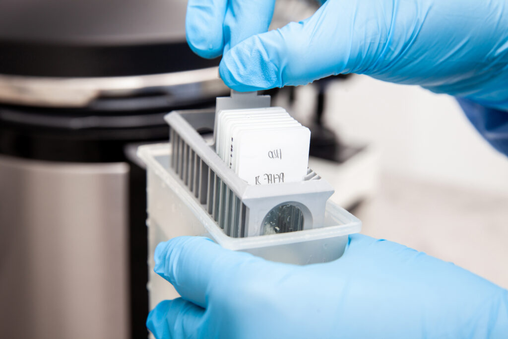
Deparaffinization removes the paraffin wax embedding the tissue, enabling access to the preserved proteins. To do this, place your FFPE tissue section in a xylene solution to dissolve the paraffin. Typically, two 10-minute xylene washes work well. Once the tissue is deparaffinized, wash it with ethanol to remove any residual xylene, and then rehydrate it with decreasing ethanol concentrations (100%, 90%, 70%, and distilled water). This step ensures that the tissue is ready for protein extraction.
Step 2: Rehydration and Buffer Preparation
After deparaffinization, the tissue should be rehydrated to prevent protein denaturation. Prepare a rehydration buffer, common Tris-HCI, or a PBS buffer, which stabilizes the proteins. Place your sample in a buffer for about 10 minutes to allow complete hydration.
Step 3: Antigen Retrieval
Antigen retrieval is a critical step in the FFPE tissue protein extraction process as it breaks the formalin-induced cross-links, freeing the proteins. Heat-induced epitope retrieval (HIER) is the most commonly used method, where samples are heated in a high-pH or low-pH buffer solution. Depending on your protocol requirements, use a microwave, pressure cooker, or water bath to heat the samples for approximately 20–30 minutes. After antigen retrieval, let the sample cool gradually to room temperature, which will reduce the risk of protein denaturation.
Protein Extraction from Tissue: 4 Key Considerations
When it comes to protein extraction from tissue, ensuring protein integrity and concentration is essential for downstream applications. Some factors to keep in mind include:
- Buffer Composition: Buffer selection should stabilize proteins without interfering with downstream analyses.
- Temperature Control: Maintaining an optimal temperature, typically 4℃, can prevent protein degradation.
- Homogenization: Mechanically disrupt tissue samples to release proteins into the solution effectively.
Protease Inhibitors: Adding protease inhibitors helps prevent protein degradation during extraction.
Protein Extraction Methods: Comparing Approaches
Several protein extraction methods exist for FFPE tissue, each with its advantages and limitations. Here are three commonly used methods:
- Heat-Induced Epitope Retrieval (HIER)
HIER is widely used for protein extraction from FFPE tissue due to its effectiveness in breaking cross-links. However, extended heating can sometimes degrade proteins, so optimization is essential. This method is ideal for labs equipped with heating devices like microwaves or pressure cookers and is compatible with immunohistochemistry (IHC) and mass spectrometry.
- Enzymatic Digestion
Enzymatic digestion uses proteases to break down proteins into peptides, making it particularly useful for mass spectrometry. However, this method may not be suitable if your study requires intact proteins. Still, it provides valuable insights into protein composition and expression levels in FFPE samples.
- Detergent-Based Methods
Detergent-based extraction involves using detergents like SDS or Triton X-100 to solubilize proteins from FFPE samples. This method can yield high protein quantities but might be incompatible with downstream applications requiring non-denatured proteins.
How to Extract Protein from FFPE Tissue: Your Step-by-Step Guide
Here’s a more detailed overview of how to extract protein from FFPE samples to achieve optimal results:
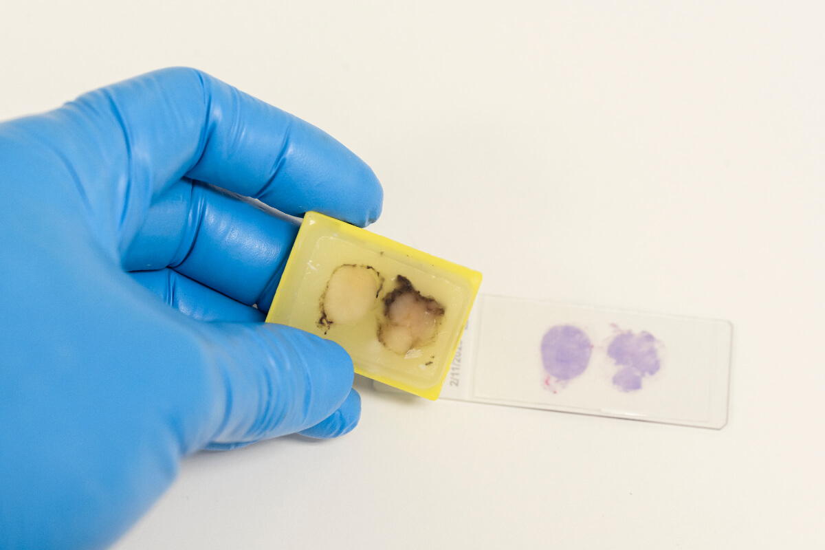
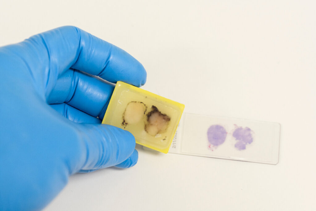
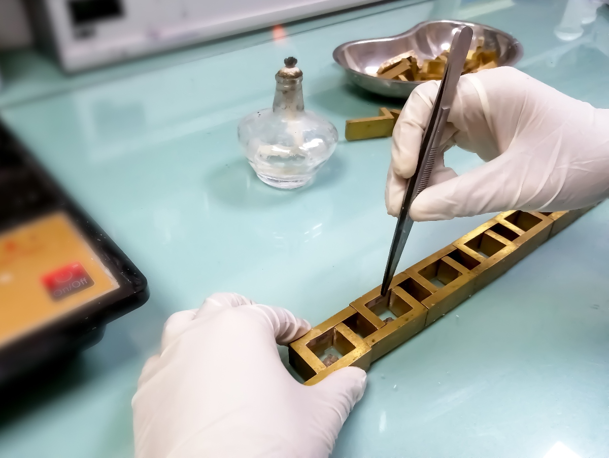
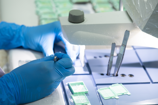
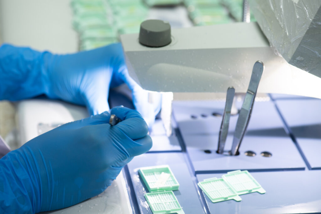
- Sample Preparation: Cut the FFPE tissue block into thin sections (typically 10-20 micrimeters thick) for better protein yield.
- Deparaffinization: Place the sections in xylene to dissolve paraffin, then wash with ethanol.
- Antigen Retrieval: Choose an appropriate pH buffer and heat the samples to reverse cross-linking.
- Lysis Buffer Addition: Add a lysis buffer, such as RIPA buffer or Tris-HCI, along with protease inhibitors. Incubate the sample on ice, using a conicator if necessary, to district tissue and release proteins.
- Centrifugation: Centrifuge the lysate at 4℃ to remove any tissue debris, collecting the supernatant that contains the extracted proteins.
- Quantification: Use a protein quantification assay, such as a BCA or Bradford assay, to determine protein concentration in the sample.
Following this protocol will maximize the yield and quality of your extracted proteins, ensuring compatibility with your downstream applications.
Optimizing FFPE Protein Extraction: Tips and Troubleshooting
Even with a solid protocol, protein extraction from FFPE tissue can sometimes yield low concentrations or degraded proteins. Here are some tips to optimize FFPE tissue protein extraction:
- Use Fresh Buffers: Old buffers can become contaminated, which may lead to protein degradation.
- Shorten Extraction Time if Possible: Prolonged extraction may lead to protein degradation. Optimize your protocol to minimize extraction time while maintaining protein yield.
- Control Temperature: FFPE protein extraction at low temperatures (4℃) can prevent protein breakdown during the process.
- Include Protease and Phosphatase Inhibitors: These inhibitors are critical for preventing protein breakdown during extraction.
Applications of FFPE Extraction Research
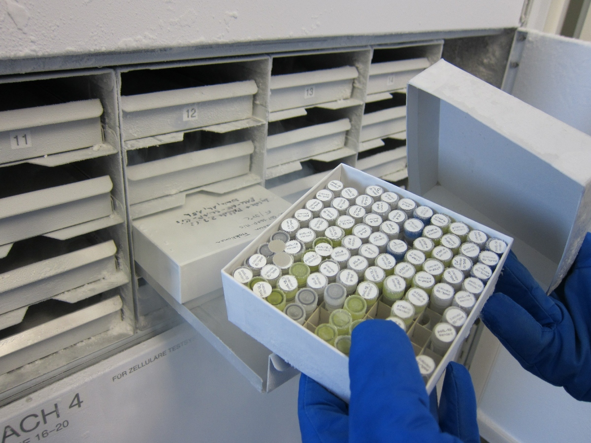
The ability to extract protein from FFPE samples opens the door for numerous research applications. FFPE protein extraction allows researchers to study historical samples, track disease progression, and perform biomarker discovery with archived tissue samples. It is widely used in fields such as oncology, pathology, and molecular biology, where preserved samples provide valuable insights into disease mechanisms and treatment responses.
Selecting the right protein extraction protocol is crucial for achieving quality results in FFPE protein extraction. The deparaffinization, antigen retrieval, and protein extraction stages must be meticulously followed to obtain high-quality proteins. Different protein extraction methods can be applied depending on the research objective, so understanding each method’s pros and cons is important.
Order Groundbreaking Quality FFPE Samples with Superior BioDiagnostics
Mastering FFPE protein extraction requires a carefully optimized protocol and suitable reagents. Whether you’re working on a biomarker, discovery, or proteomic analysis, extracting proteins from FFPE tissue samples can yield valuable data. Superior BioDiagnostics is committed to providing top-quality FFPE samples for your research, including normal, malignant, and disease-state breast, cervical, muscular, and more. Our expertise in biobanking ensures reliable samples to enhance your research outcomes. Contact Superior BioDiagnostics today and order reliable, high-quality FFPE samples to support your protein extraction work.