How to Prepare Tissue Samples For Microscope Slides: A Step-By-Step Guide
Histology technicians play a significant role in biological research. Without these scientific experts, we wouldn’t have tissue slides carrying thin sections of biospecimens ready for microscopic analysis. Tissue samples prepared for microscope slides have radically impacted science; researchers can study cellular structures, disease traits, and the effects of various treatments on specimens. Pathologists can accurately diagnose diseases such as cancer, inflammatory conditions, and bacterial infections. Without histology technicians, we wouldn’t have tissue samples properly prepared for microscopic analysis.
You may ask the question, “How do histology technicians prepare tissue samples for microscope slides?” In this blog, we share the 5 steps it takes to prepare human tissue samples for microscopic study. Whether you’re an experienced histology technician or simply curious about what goes into microscopic examination, this article is your guide to preparing tissue samples for microscope slides. From the fixation of human tissues to various staining techniques, you’ll walk away knowing exactly how to prepare biospecimens for microscopic research purposes.
What You Need for Tissue Sample Preparation
Before you get started, there is an essential list of tools you will need for tissue sample preparation. The supplies list depends on the tissue type and what you’re using the biospecimen for. However, here are some staples you’ll most likely need when you prepare tissue samples for microscope slides:
- Fixatives (e.g., formalin, glutaraldehyde, etc.)
- Processing solutions (e.g., xylene, ethanol, and other solutions as needed)
- Embedding medium
- Microtome or cryostat
- Staining agents
- Water bath
- High-quality slides and coverslips
- Slide labels
- Slide storage boxes
Remember that the supplies you need for tissue preparation vary depending on the tissue type, staining method, and studies to be conducted. Ensure that you follow the protocols and safety guidelines of your laboratory or scientific institution when prepping tissue samples for microscopic analysis.
Tissue Sample Preparation for Microscope Slides: 5 Simple Steps You Can Follow
Once you’ve gathered all the necessary material, you’re ready to prepare tissue samples for microscope slides. Whether you’re new to it or not, you likely understand how large of an undertaking tissue sample preparation is. That’s why we’re here to give you a complete step-by-step guide to preparing tissue samples for microscopic study. Below are the 5 steps you can follow to prepare tissue samples for microscope slides:
Fix the biospecimens
After obtaining the fresh tissue sample, it must be fixated, whether that’s through freezing or chemical fixation. It’s important to start the fixation process immediately after collecting the biospecimen, ensuring that it holds its original structures and molecular makeup. In turn, you’ll get the best results! Fixation is critical for the rest of the tissue sample preparation. Fixation preserves the chemical composition of the biospecimen, securing and hardening the sample to initiate easy sectioning. Below are the two primary types of fixation that researchers use for tissue sample preparation:
- Fixative Solution: Researchers immerse biospecimens in a chemical fixative solution for 6–24 hours shortly after collection. Neutral Buffered Formalin or Paraffin-formalin are popular, effective solutions scientists and laboratories use for chemical fixation. Fixative solutions must penetrate every part of the biospecimen, preserving the sample and preparing it for microscopic analysis.
- Freezing: Scientists submerge the tissue samples in a tissue-freezing medium, which is then immersed in liquid nitrogen. Freezing biospecimens is an alternative to the fixative solution method and is preferred by researchers who need an immediate diagnosis.
Fixing human tissue samples, whether by freezing or using a chemical fixative, assists in preserving specimens and preventing degradation. Once you fixate the biospecimens, they’re ready to be processed.
Process the tissue samples
Tissue processing can be performed with an automated machine or by hand, preparing the biospecimen for sectioning. Here are three general steps involved in tissue processing:
- Dehydration: Before converting the biospecimen into a solid form appropriate for sectioning, it must undergo dehydration. Ethanol is a popular agent used for the dehydration process. Ethanol removes water from the tissue sample and hardens it for microscopic use. Researchers immerse the specimen in multiple ethanol solutions of increasing intensity, ensuring that the tissue is free of water and formalin.
- Clearing: After the submergence of ethanol and before the embedment of wax, the tissue sample needs an in-between “clearing” medium that is compatible with both paraffin wax and ethanol. Xylene is a popular solvent that rids tissue of ethanol and prepares it for infiltration of wax.
- Embedding: Using an embedding center, the biospecimen is placed into a mold of molten wax (typically paraffin wax), forming what is called a “block” ready for sectioning. The newly created block is then cooled and prepared for thin-section cutting. Embedding also preserves the cellular structure of a tissue sample, making it more compatible with succeeding tissue sample preparation steps.
Processing helps preserve the integrity of tissue samples, leading to more accurate analysis and improving scientific diagnostics. Processed biospecimens also absorb stains more effectively and can be stored long-term in a biobank if needed. In summary, proper processing sets you up for success during the rest of the tissue sample preparation procedure.
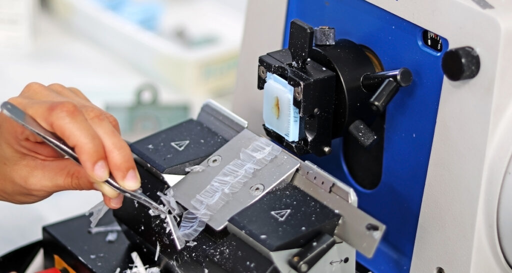
Cut the biospecimens into sections
Now, your biospecimen is ready to be cut into sections on a microtome. First, the wax is removed from the surface of a block, exposing the tissue sample. Using a microtome, the biospecimen is sliced into sections no more than 4–5 micrometers. A microtome can cut continuously, creating a “ribbon” of tissue sections that are perfect for microscopy. If the sample is frozen, you’ll use a cryostat to cut it into tissue ribbons. These specimen ribbons can then be placed in a warm water bath to flatten. From here, tissue samples are easily collected for staining and examination on a microscopic slide.
Stain the samples
Staining specimens is a vital element of tissue sample preparation. While there are numerous staining techniques, we’ll highlight a few of the most common methods below:
- Hematoxylin and Eosin: The use of Hematoxylin and Eosin (H&E) is the most widely used staining technique for pathologists. Hematoxylin is a dye that stains acidic structures, while eosin is a counterstain done after hematoxylin that marks the sample’s basic structures. The result? Cell nuclei are often colored blue/purple, and other cellular structures that attract eosin (eosinophilic structures) are stained with a pink/red hue. RNA in ribosomes or the rough endoplasmic reticulum, for instance, would be stained blue, while the cytoplasm would be colored pink.
- Gram Staining: Gram staining is used primarily to differentiate bacterial species according to the physical and chemical components of their cell walls. With the help of a gram stain, differing bacteria will change one of two different color sets (purple to blue or pink to red). Bacteria are then labeled as “gram-positive” or “gram-negative” according to their coloring. Gram-positive bacteria contain a hefty layer of peptidoglycan, making them appear purple. Gram-negative bacteria, on the other hand, hold a thin layer of peptidoglycan and other lipids in the cell wall, which washes out the violet in the decolorization process and stains them pink. Gram staining aids in diagnosing bacterial infections and the types of bacteria causing the illness.
- Masson’s Trichrome: Masson’s trichrome staining is a popular method that produces multicolor results on biospecimens. In a trichrome staining procedure, collagen is stained blue, muscle tissue is colored red, cytoplasm is dyed pink, and nuclei have a dark brown tint. The trichrome technique can distinguish collagen from muscle and identify pulmonary fibrosis, cardiac fibrosis, chronic kidney disease, muscular dystrophy, and various tumors of muscle origin.
Staining tissue samples enhances scientific studies by identifying a tissue sample’s components such as proteins, lipids, and carbohydrates. Varied stains can also aid in diagnosing diseases, differentiating normal and abnormal specimens, and detecting the presence and location of particular proteins. Overall, staining helps histologists better understand the minuscule structure and function of normal, malignant, and disease-state tissues.
Mount the tissue sample sections for microscopic examination
After the biospecimen has been stained, the tissue sample section is ready to be mounted between a slide and coverslip, ensuring that it’s secure and ready for microscopic examination. Outlined below are the steps of mounting a slide:
- Apply a single drop of an aqueous-based or resinous mounting medium onto the tissue section.
- Hold the coverslip at a 45° angle and allow the drop to spread to the edge of the slip.
- Gently let go of the slip so it covers the tissue section, allowing the medium to spread slowly and cover the biospecimen completely.
It’s important to utilize a clearing agent that’s compatible with the mounting medium, preventing issues in the mounting stage. It’s also necessary to label each slide with the patient ID, tissue type, and date. After mounting your biospecimen, it’s ready to be stored for observation under a microscope! Store tissue samples in a cool, dark place to prevent the fading of stains.
Receive the Finest Tissue Samples Prepared for Microscopic Study
Do you want to skip the tissue sample preparation process and invest in high-quality biospecimens prepared exclusively for microscopic analysis? Superior BioDiagnostics has everything you need and more. Our biorepository is stocked with thousands of normal, malignant, and disease-state FFPE tissue samples ready for your research purposes. We provide biospecimens from just about every anatomical site in the form of blocks, slides, and sections.
We collect tissue samples that are 100% US-procured and processed, ensuring you receive the purest biospecimens for microscopic analysis. Superior Biobank’s team can also include data on the tissue sample’s tumor type (if applicable), TNM stage, histologic grade, and donor demographics to improve your discoveries. Contact Superior Biobank to order pre-prepared tissue samples for your microscopic study.