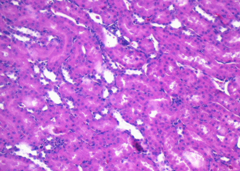
Optimizing Immunostaining Techniques for FFPE Tissue
Immunostaining is a vital technique in molecular biology and pathology, used to identify specific proteins, antigens, or other biomolecules in tissue samples. Its application is particularly important for studying disease states, diagnosing various cancers, and researching pathophysiological mechanisms. Formalin-fixed paraffin-embedded (FFPE) tissue samples are commonly used in immunostaining, offering advantages such as preservation of tissue structure and antigenicity. Let’s explore the nuances of immunostaining protocols to understand how to select the right immunostains to ensure proper use of antibodies for optimal results.

What is Immunostaining?
Immunostaining, also known as immunohistochemistry (IHC) or immunocytochemistry (ICC), involves using antibodies to detect the presence of specific antigens within a tissue sample. The process is based on the specificity of antibodies binding to particular proteins or other molecules in the tissue, which is then visualized using chromogenic or fluorescent detection methods. FFPE tissue, in particular, is often used for this technique due to its ability to preserve tissue morphology over long periods.
The FFPE process involves fixing tissue samples in formalin and embedding them in paraffin wax. This technique preserves the tissue’s morphology and molecular integrity, making it ideal for long-term storage and subsequent immunostaining procedures.
The Immunostaining Protocol
The immunostaining process begins with a careful preparation of FFPE tissue, which is sectioned into thin slices and mounted on glass slides. Before staining can occur, these sections are deparaffinized to remove the paraffin wax and rehydrated to prepare the tissue for aqueous solutions. One critical step in the process is antigen retrieval, which involves exposing the tissue to heat and a retrieval buffer to unmask antigens that may have been masked during formalin fixation. This ensures antibodies can bind effectively to their target antigens.
A blocking agent is applied to minimize background noise, which helps reduce the non-specific binding of the antibodies after immunostaining. This is followed by the application of a primary antibody, which is selected based on the antigen of interest. For instance, researchers investigating HPV-related cancers often use a p16 immunostain, while those studying gastric conditions might choose an immunostain for Helicobacter pylori. Once the primary antibody binds to its target, a secondary antibody conjugated with a detection marker is introduced to amplify the signal.
Detection methods vary depending on the study’s needs. Some laboratories use colorimetric systems, where a visible dye indicates the presence of the antigen, while others use fluorescence for more detailed imaging. Finally, counterstaining highlights the overall tissue morphology, making it easier to interpret results in the context of the tissue structure.
One additional consideration is the temperature and duration of antigen retrieval. Over-retrieval can cause tissue damage or loss of antigenicity, while under-retrieval may result in weak or absent staining. Adjusting retrieval conditions for specific antibodies can significantly enhance outcomes. Similarly, using high-quality mounting mediums and coverslips ensures that the final slide is preserved for analysis without compromising the immunostaining results.
Comparing SMA vs. MSA or CMV Immunostaining Applications
Immunostains are used across various applications, each tailored to specific research or diagnostic needs. For example, smooth muscle actin (SMA) and muscle-specific actin (MSA) immunostains help distinguish between smooth and strained muscle tissues, which is critical in diagnosing various muscle-related diseases. Similarly, p16 immunostains serve as surrogate markers for HPV infection and are widely used in detecting HPV-related cancers.
In infectious disease research, immunostains are vital for detecting pathogens. An immunostain for Helicobacter pylori is commonly employed to identify the bacterium in gastric tissues, helping diagnose and monitor H. pylori infections. Cytomegalovirus (CMV) immunostains are similarly invaluable in identifying viral antigens in tissues, particularly in transplant or immunocompromised patients.
When interpreting results, it’s important to note that a negative p16 immunostain result might mean the absence of HPV-related lesions, offering insights into whether a particular cancer is HPS-driven. Each type of immunostain provides critical information, enabling pathologists and researchers to make informed decisions about diagnoses and treatments.
The specificity of immunostains also enables pathologists to differentiate between closely related diseases. For example, in distinguishing smooth muscle tumors, SMA and MSA immunostains provide complementary data that can influence diagnostic accuracy. Moreover, advancements in multiplex immunostaining now allow simultaneous detection of multiple antigens, opening doors for more comprehensive insights into complex tissue samples.
Challenges and Optimization in Immunostaining FFPE Tissue
Immunostaining FFPE tissue samples can present unique challenges. Formalin fixation, while excellent for preserving tissue morphology, may mask certain antigens, requiring careful optimization of antigen retrieval methods. Choosing the right antibodies is equally important, as antibody specificity and sensitivity significantly impact results. Non-specific binding and background staining are common issues but can be minimized by adjusting blocking agents and optimizing antibody concentrations after immunostaining.
To ensure the best outcome, it’s essential to validate antibodies for FFPE use and tailor protocols to the specific tissue and antigen. Incorporating control samples, both positive and negative, can help confirm the accuracy and specificity of the immunostaining process.
Another key optimization strategy is using digital pathology tools. These systems can analyze immunostained slides with greater precision, reducing inter-observer variability. Automated imaging solutions also facilitate large-scale studies, ensuring consistent results across multiple samples. Additionally, the introduction of recombinant antibodies has enhanced the reproducibility of immunostaining by eliminating batch-to-batch variability found in traditional antibodies.
Order Top-Quality Samples For Your Research
At Superior BioDiagnostics, we understand the complexities of immunostaining and the importance of high-quality FFPE tissue samples in achieving reliable results. Our biobank offers a diverse array of FFPE tissue blocks, sections, and slides, including normal and disease-state breast, cervical, lung, muscle tissues, and more. These samples are meticulously processed to maintain their structural integrity, making them ideal for immunostaining and other molecular analyses.
Whether you’re investigating the expression of p16 in cervical cancer, testing immunostains for H. pylori in gastric tissues, or exploring novel biomarkers, our specimens provide the reliability and precision needed for groundbreaking discoveries. Superior BioDiagnostics is committed to supporting your research with next-day shipping and comprehensive data to accelerate your findings.
Explore our selection of FFPE tissue samples today. With unmatched quality and global shipping, we’re here to help you reach your next breakthrough. Contact Superior BioDiagnostics to place your order and access the resources you need to drive innovation forward.
