Digital Pathology Today: Trends Defining the Future
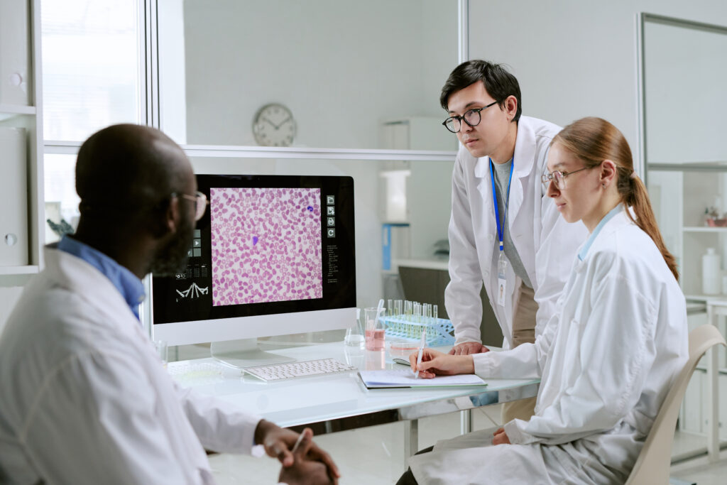
Digital pathology has moved far beyond early experimentation and pilot programs. Today, laboratories, pharmaceutical companies, and research institutions worldwide are integrating digital tools into everyday pathology workflows. High-resolution whole slide imaging, AI-assisted diagnostics, and advanced tissue analytics are transforming how pathologists review, interpret, and share data.
When discussing digital pathology today, it’s clear that the field is entering a new phase of growth. Instead of focusing solely on digitizing slides, organizations are building entire ecosystems that combine imaging, analytics, molecular data, and collaboration platforms.
These developments are helping shape the future of digital pathology, where faster diagnostics, more scalable research, and improved biomarker discovery become standard practice across healthcare and life sciences.
In this article, we’ll explore the key trends driving digital pathology today and what they reveal about the next stage of this rapidly evolving field.
The Expansion of Digital Pathology in Clinical and Research Settings
One of the most significant developments in digital pathology today is the rapid expansion of digital workflows in both clinical diagnostics and biomedical research.
Traditionally, pathology relied on glass slides viewed under a microscope. While this approach remains foundational, digital scanning technologies now allow slides to be converted into high-resolution digital images. These digital slides can then be analyzed, shared, and archived far more efficiently than traditional slides.
For research teams and biotech companies, this shift unlocks major advantages. Digital slides enable:
- Remote collaboration between pathologists and researchers
- Faster access to tissue data across multiple locations
- Improved data integration with genomic and molecular datasets
Many organizations working in digital pathology also rely on standardized tissue materials to support research and development. For example, FFPE samples remain one of the most widely used formats for histology and biomarker studies. Researchers interested in understanding how preserved samples support digital analysis can explore more about FFPE tissue and its role in modern pathology workflows.
As digital imaging becomes more accessible, the use of digital slides in research pipelines will continue to expand.
Whole Slide Imaging and Data Infrastructure
Whole slide imaging (WSI) is one of the foundational technologies enabling digital pathology today.
WSI scanners capture high-resolution images of entire microscope slides, producing digital files that can be stored, analyzed, and shared across systems. These digital pathology slides enable pathologists to review tissue samples remotely while maintaining the level of detail required for accurate diagnosis.
For laboratories transitioning to digital workflows, managing digital slide data is a key challenge. High-resolution slide files can be extremely large, requiring robust storage, processing, and viewing systems.
Organizations adopting digital pathology solutions often build infrastructure that supports:
- Secure image storage
- AI-assisted analysis tools
- Collaborative review platforms
- Integration with laboratory information systems (LIS)
Researchers seeking to understand the importance of digital pathology slides can learn more about their role in modern diagnostic workflows.
As computing infrastructure improves, these systems are becoming more scalable and accessible to laboratories of all sizes.
Artificial Intelligence Is Transforming Pathology Workflows
Artificial intelligence is one of the most influential drivers shaping the future of digital pathology.
AI tools are increasingly being developed to assist pathologists with tasks such as:
- Identifying tumor regions in tissue samples
- Detecting metastases in lymph nodes
- Quantifying biomarker expression
- Automating routine image analysis tasks
Rather than replacing pathologists, these technologies are designed to augment human expertise by providing faster, data-driven insights.
For example, several AI-powered pathology platforms can now analyze thousands of digital slides and identify patterns that may not be immediately visible during manual review.
Readers interested in exploring how machine learning is being applied in this space can learn more about AI in digital pathology and its growing role in diagnostic innovation.
As AI algorithms continue to improve, their integration into digital pathology platforms will likely become a standard component of modern diagnostic workflows.
Recent Industry Developments Highlight Momentum in Digital Pathology
The momentum behind digital pathology today is also visible in recent industry developments.
A digital pathology roundup in September 2025 highlighted major funding rounds, regulatory approvals, and strategic partnerships that signal continued investment in this field.
Three key developments illustrate how rapidly the sector is evolving:
1. Major Funding for AI and Digital Pathology Platforms
Cyted Health raised €44 million in Series B funding led by EQT Life Sciences to support expansion into the United States and expand testing capabilities beyond its EndoSign platform.
Similarly, StratifAI secured €12.5 million in funding to validate its multimodal AI platform, Polaris. This platform integrates H&E histology images, RNA sequencing data, and clinical outcomes to generate spatial insights that could improve prognostic and predictive models.
These investments highlight growing confidence in digital pathology technologies and their potential to reshape diagnostics.
2. Regulatory Approvals for AI-Based Diagnostic Tools
Several companies also received important regulatory approvals in 2025.
Aiforia obtained CE-IVD approval for an AI-based tool designed to detect lymph node metastases. According to the company, the technology could reduce cancer staging time by up to 40%.
Primaa also secured CE-IVDR approval for Cleo Breast, a platform designed to automate biomarker detection on biopsy and surgical specimens, improving diagnostic efficiency.
These regulatory milestones demonstrate how digital pathology tools are moving from experimental technologies into real clinical applications.
Partnerships Expanding the Digital Pathology Ecosystem
Strategic collaborations are another major trend defining digital pathology today.
Lunit partnered with Leica Biosystems to distribute its AI tool through the Aperio AI Store, while also working with Agilent to develop AI-powered companion diagnostics.
PathAI has expanded its AISight Dx platform through partnerships with Mindpeak, Stratipath, and Primaa, integrating multiple CE-IVD tools for breast and lung pathology.
Meanwhile, Proscia introduced a platform called Aperture designed to convert diagnostic pathology data into real-time insights for biomarker development and regulatory submissions.
Together, these developments illustrate how digital pathology platforms are becoming interconnected ecosystems rather than isolated tools.
Digital Pathology and the Future of Biomarker Discovery
One of the most promising aspects of the future of digital pathology lies in its potential to accelerate biomarker discovery.
Digital pathology allows researchers to analyze tissue samples at scale, combining morphological features with genomic, proteomic, and clinical data. This integration is particularly valuable in oncology research, where understanding tumor microenvironments and spatial biology is essential.
Tissue-based research remains a critical component of biomarker development. Researchers studying disease progression often rely on carefully characterized tissue samples, including both malignant and normal tissue types.
For example, cancer researchers may use curated malignant tissue samples to study tumor morphology and therapeutic response.
Comparative analysis often requires reference material, such as normal tissue samples used to establish baseline biological patterns.
By combining high-quality tissue resources with digital imaging and analytics, researchers can generate insights that support drug discovery and personalized medicine.
Digital Pathology Is Enabling Global Collaboration
Another key advantage of digital pathology is the ability to collaborate across geographic boundaries.
With digital slides and cloud-based platforms, pathologists can review the same tissue samples from different locations. This capability is particularly valuable for:
- Multi-site clinical trials
- Rare disease research
- Specialist consultation and second opinions
In traditional pathology workflows, physical slides must be shipped between laboratories, a process that can take days or even weeks.Digital pathology eliminates this delay, allowing experts to review cases almost instantly.
As digital infrastructure continues to improve, global collaboration will likely become a defining feature of the future of digital pathology.
The Role of High-Quality Tissue Samples in Digital Pathology
Even as digital tools transform pathology workflows, one constant remains: the need for high-quality biological samples.
Digital imaging and AI analysis are only as reliable as the underlying tissue data they analyze. For research institutions, pharmaceutical companies, and diagnostic developers, access to well-characterized tissue samples is essential.
Organizations conducting digital pathology research often require a reliable source of tissue samples for imaging, biomarker validation, and algorithm training.
Superior BioDiagnostics provides a range of research-ready FFPE tissue products designed to support these workflows.
For teams ready to begin a project or obtain research materials, ordering directly through the platform simplifies the process.
What Comes Next for Digital Pathology?
Looking ahead, the future of digital pathology will likely be shaped by continued convergence between imaging, artificial intelligence, and molecular data.
Some of the developments expected in the coming years include:
- Expanded use of AI-assisted diagnostics
- Integration of pathology data with genomic sequencing
- Improved regulatory pathways for digital diagnostic tools
- Greater use of digital pathology in clinical trials
At the same time, digital pathology will continue to rely on foundational elements such as standardized tissue samples, high-resolution imaging, and expert interpretation.
The technologies and partnerships emerging today suggest that pathology is moving toward a more connected, data-driven future, one in which digital tools enhance human expertise rather than replace it.
For laboratories, researchers, and biotech innovators, understanding digital pathology today is the key to preparing for what comes next.