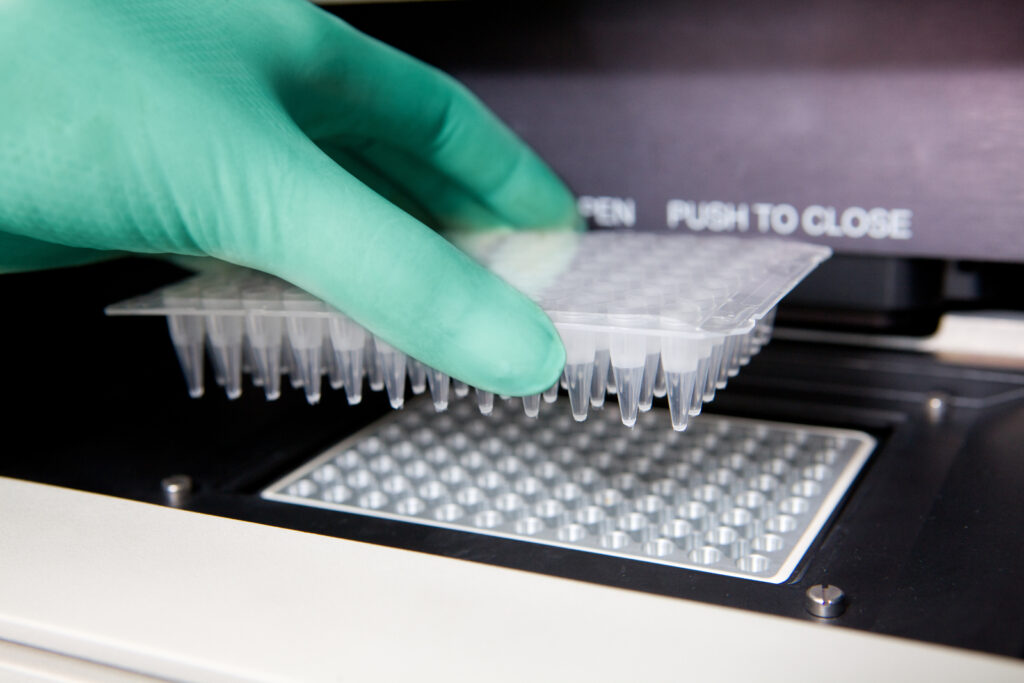
PCR, qPCR, and RT-PCR: Differences Between Real-Time, Reverse Transcription, and Traditional PCR
Polymerase Chain Reaction (PCR) and its variations, qPCR (quantitative PCR), and RT-PCR (Reverse Transcription PCR) are essential techniques in molecular biology. While all three methods involve DNA amplification, they serve different purposes—traditional PCR amplifies DNA, qPCR allows real-time quantification, and RT-PCR enables RNA detection by converting it into DNA. In this blog, we’ll break down their differences and applications in research.

What is PCR?
Polymerase Chain Reaction is a technique for amplifying or producing multiple copies of a short DNA or gene stretch. It is commonly used in molecular biology and biotechnology, allowing researchers to make billions of copies of a specified DNA for research and diagnostic purposes. Traditional PCR involves three steps: denaturation, annealing, and extension of primers.
It requires two primer sets that are complementary to both ends of the DNA template and a thermostable DNA polymerase. The polymerase cycle is repeated multiple times to generate numerous copies of the DNA segment.
Here’s a breakdown of the 3 steps of PCR:
- Denaturation (95°C): During this step, the reaction mixture is heated to a high temperature, typically around 94–98°C. This heat breaks the hydrogen bonds between the base pairs in the double-stranded DNA, causing it to separate into two single strands. The separation of DNA strands is crucial because it allows primers to bind to the template in the next step.
- Annealing (50–65°C): The temperature is then lowered to 50–65°C, allowing short primers to attach (anneal) to complementary sequences on the single-stranded DNA. Primers are short DNA sequences that act as starting points for DNA synthesis. The temperature used in this step depends on the primers’ melting temperatures, ensuring specific and stable binding.
- Extension (72°C): The reaction temperature is then raised to 72°C, the optimal working temperature for Taq polymerase, a heat-stable enzyme. This enzyme adds nucleotides (A, T, C, G) to the primers, extending the DNA strand in the 5′ to 3′ direction. As a result, two identical copies of the original DNA sequence are formed.
This process is repeated 30–40 times, exponentially increasing the number of DNA copies with every cycle.
What is qPCR?
Quantitative PCR, or qPCR, is also known as real-time PCR. It’s a technique that uses fluorescent reporter molecules to measure the amount of RNA or DNA in a tissue sample. qPCR gives an additional quantitative analysis of the DNA or RNA in RT-qPCR, which we’ll explore further below.
Researchers use real-time PCR to measure gene expression, viral load, and SNP detection. It’s also a vital tool for genetic testing, pathogen detection, and disease research. In qPCR or real-time PCR, the amplification of DNA is monitored in real-time using fluorescence-based detection.
There are two primary methods for detecting PCR products in real time:
- Dye-Based Detection (SYBR Green): Uses SYBR Green, a fluorescent dye that binds specifically to double-stranded DNA. As DNA amplifies, the fluorescence signal increases proportionally to the DNA concentration. Dye-based detection is a simple and cost-effective method, but it may detect non-specific products such as primer dimers.
- Probe-Based Detection (TaqMan, Molecular Beacons, Scorpion Probes): Uses fluorescently labeled probes that bind specifically to the target DNA sequence. It’s more specific than dye-based detection as it only detects the target sequence, reducing the background signal. There are 3 types of probe-based methods:
- TaqMan Probes: This probe has a fluorophore and quencher. Fluorescence is released when DNA polymerase cleaves the probe during amplification.
- Molecular Beacons: A hairpin-shaped probe that fluoresces when it binds to the target DNA.
- Scorpion Probes: Similar to molecular beacons, they are integrated into the primer for faster and more efficient detection.
Both methods allow DNA quantification in real-time, but probe-based detection offers greater specificity and accuracy, making it preferable for applications like gene expression analysis and pathogen detection.
What is RT-PCR or Reverse Transcription PCR?
RT-PCR (Reverse Transcription PCR) is a modified form of PCR that amplifies RNA by first converting it into complementary DNA (cDNA) using the enzyme reverse transcriptase. This allows the detection of RNA-based genetic material, making it useful for studying gene expression and detecting RNA viruses like SARS-CoV-2 (COVID-19).
RT-PCR consists of two steps: first, the reverse transcription process converts RNA into complementary DNA (cDNA), and second, the polymerase chain reaction amplifies the desired DNA sequence. RT-PCR is used to detect RNA in a sample, but it does not measure RNA quantity. To quantify RNA levels, RT-qPCR (quantitative reverse transcription PCR) is used. RT-qPCR combines RT-PCR with qPCR, making it highly useful for quantitative viral RNA and gene expression analysis.
Differences Between PCR, qPCR, and Reverse Transcription PCR
Both qPCR and real-time PCR are advanced methods of polymerase chain reaction. While RT-PCR is used to detect and amplify cDNA, qPCR provides quicker, more detailed real-time results and is utilized to quantify nucleic acids.
Here’s a detailed chart to understand the primary differences between PCR, qPCR, and reverse transcription PCR:
PCR | qPCR | RT-PCR |
| Used to analyze a short stretch of DNA by amplification | Advanced PCR method used to amplify and quantify DNA | Used to detect and amplify RNA by converting it into DNA |
| Primer is used for polymerization | Primer, as well as fluorescent probes or dyes, are used | Reverse transcriptase enzyme is used to synthesize complementary DNA (cDNA) from RNA |
| Results are investigated by gel electrophoresis | Fluorescence emitted by dye or probe is recorded during PCR | Results are investigated by gel electrophoresis |
| Data is recorded at the end of the process | Data is recorded during the amplification process at the exponential phase | Data is recorded at the end of the process |
| Ethidium bromide is used to stain DNA fragments | Fluorescent dyes or DNA probes labeled with a fluorescent reporter are used | Ethidium bromide or fluorescent dyes can be used to stain DNA fragments |
| Low-resolution technique | High-resolution technique | Low-resolution technique (unless combined with qPCR) |
| Distinct bands of various DNA fragments are seen on agarose gel | Different peaks related to DNA fragments seen during qPCR | Distinct bands of DNA fragments seen on agarose gel |
| It takes more time to generate a result | Requires less time to deliver results | It takes more time than qPCR due to an additional reverse transcription step |
| Detects the presence or absence of DNA and gene mutations, and it amplifies templates for sequencing | Quantifies DNA, analyzes gene expression, detects pathogens, and quantifies and identifies mutations | Detects RNA viruses (e.g., COVID-19), analyzes gene expression, and studies mRNA levels |
While PCR is primarily used for DNA amplification, qPCR enables real-time quantification, and RT-PCR allows RNA detection by converting it into DNA. Understanding these differences is essential for selecting the appropriate method based on research or diagnostic needs.
Order FFPE Tissues for Reliable PCR, qPCR, and RT-PCR Testing
Using high-quality FFPE tissues is essential for accurate and reliable results when performing PCR, qPCR, or RT-PCR. Superior BioDiagnostics offers a diverse selection of normal, malignant, and disease-state FFPE tissues from nearly every anatomical site, ensuring you get the right samples for your research and diagnostic needs. Whether you’re analyzing gene expression, detecting pathogens, or conducting molecular testing, our high-quality FFPE tissues provide the consistency and precision you can trust. Order today and advance your PCR-based studies with confidence.
